Veterinary Refresher Course: Foot Lameness
The Horse’s Foot
Anatomy: What is the foot?
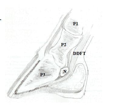
Bones: The horse’s foot includes the structures below the fetlock, sometimes called the “ankle”. Similar to our third, or middle, finger anatomically, the foot is a remarkable complicated structure. There are 3 bones that make up the structural framework of the foot: the phalanges.
- Phalanx 1 (P1) = Proximal Phalanx = Long Pastern Bone
- Phalanx 2 (P2) = Middle Phalanx = Short Pastern Bone
- Phalanx 3 (P3) = Distal Phalanx = Coffin Bone
Additionally, located in the foot is the navicular bone (N). This boat-shaped bone is a sesamoid bone. This is a type of bone that is found where a tendon passes over a joint. There are several in different locations throughout the body.
Joints: there are two joints in the horse’s foot: Proximally (closer to the horses body) is the proximal interphalangeal joint (PIJ), AKA the pastern joint. This joint is between P1 and P2. Distally is the distal inter-phalangeal joint (DIJ), AKA coffin joint. This joint is between P2, P3 and the navicular bone.
Ligaments: collateral ligaments help to stabilize the joints. There are short and long versions on the inside and outside of each of the joints. Additionally, ligaments help to hold the navicular bone in place against the coffin and short pastern bones.
Tendons: Allow the horse to move its food. The extensor tendon across the front lifts the toe. The flexor tendons along the back lift the heels and flex the coffin and pastern joints.
Navicular bursa: A fluid filled sac that sits between the navicular bone and the deep digital flexor ten-don, providing cushioning
Ungual Cartilages: Shock absorbers in the hoof that are attached to the coffin bone
Hoof: Additional structures include the hoof capsule, sole and digital cushion (shock absorber)
Diagnostics:
Exam: our exam always includes palpation, passive range of motion flexion and hoof testing. Hoof testers are used to find sore spots around the sole, frog and across the heels.
Regional anesthesia: AKA “blocking”. Specific structures or regions are numbed and the horse is re-evaluated to determine what areas and/or structures could be causing a lameness.
It is possible to numb either the hoof or the pastern and the hoof.
Intra-articular anesthesia: blocking joints. It is possible to block the pastern joint, coffin joint and navicular bursa.
Radiographs: Compress a 3D object into a 2D image. This means multiple views of each structure are required for full assessment of a structure. Most commonly, a lateral (side to side) and dorsopal-mar/dorsoplantar (front to back) are performed to assess for rotation of the coffin bone, medial to lateral (inside to outside) balance, toe length, sole depth, etc. Two additional views are used to assess the navicular bone, the dorsopalmar 60o oblique and the skyline views.
Ultrasound: Used to visualize “soft tissue” structures, including ligaments and tendons. Joint capsules are not easily assessed unless there is an abnormality present. Ultrasound uses sound waves to create images and is unable to image through bone, air or the hoof capsule. Ultrasound is very useful, but limited primarily to the pastern region.
Further imaging: For a complete diagnosis, some horses require MRI, CT or a bone scan. All of these require referral.
Lameness:
Approximately 90% of lameness originates in the foot. There are a lot of small structures that can be affected. Lameness is graded on a scale of 1-5, depending on severity. This allows description of the lame-ness in your horse’s medical record and allows veterinarians to speak accurately to each other when describing a lameness. Some signs of lameness include: changes in stride length, “head nodding” while walking and/or trotting, unwillingness to bear weight on a limb. At times the only complaint is a horse that does not move or perform as they had previously.
Hoof Abscesses
Sometimes called “gravels”. Hoof abscesses often present as a 5/5, or non-weight bearing, lameness. Abscesses can be subsolar (under the sole) or in the heel region. Some abscesses can erupt from the coronary band. Signs include acute (sudden) extreme lameness of one limb. This is often combined with increased digital pulses (blood flow into the foot), heat in the foot and/or soreness to hoof testers. Treatment requires opening of the abscess to allow drainage. Think of a hoof abscess like a pimple—it is a pocket of puss that is under a lot of pressure and therefore very painful. Treatments include soaking—often in warm water and ep-some salts; poultice wraps—sometimes with MagnaPoultice®, ichthammol and/or Animalintex® poultice pads; cutting an abscess open. In rare cases, sometimes we will block a foot to encourage a horse to walk on the foot to encourage the abscess to “pop”.
Palmar/Plantar Heel Pain
Also called “Navicular disease” or “Navicular syndrome”. The advent of MRI has shown that this syn-drome can be caused by many different problems, hence the name changes. There are numerous causes, in-cluding deep digital flexor tendonitis, navicular bursitis, inflammation of the navicular bone, coffin joint arthri-tis/pain, collateral ligament desmitis, other injured structures and/or “-itis’s”. Treatment is variable, depending on the underlying cause of the lameness. This includes shoeing changes—pads, wedges, special shoes; injec-tions—coffin joint, navicular bursa; pain relief—NSAIDs (ie Bute, Banamine, Naproxen, etc), sometimes isoxuprine is used (very case dependent). As always, all treatment recommendations are case dependent, and this includes rest.
Ringbone
Extra bone formation along the long and/or short pastern bone(s). Most often due to instability near the joint and/or ligamentous damage.
Articular v Non-Articular: involving a joint v no joint involvement
High v Low Ringbone: High—pastern joint disease
Low—coffin joint disease, sometimes also called buttress foot if there is appreciable deformity of the hoof capsule and/or area just above the coronary band
Treatment also involves shoeing recommendations, medications such as NSAIDs and injections. In some cases, fusions of the pastern joint is recommended to increase the comfort level of a horse with ex-tremely painful ringbone.
Sidebone
Calcification of the previously mentioned ungula cartilages. Thought to be cause by instability in the hoof and sometimes related to concussion. Conformation and breed may also influence the likelihood of sidebone formation. Lameness is typically rare. Treatment is only needed if the horse is painful.
Hoof Foreign Bodies
You should never remove a foreign body prior to consulting with your veterinarian. Most foreign bod-ies are metal and a radiograph (x-ray) should always be taken to assess what structures are affected by the for-eign body prior to its removal. Based on this, necessary treatment can be determined. Treatment is variable based on affected structures, and includes NSAIDs and antibiotic therapy. Sometimes regional limb perfusion (local injection of antibiotics) and/or surgery can be required.
Overall Hoof Health:
Farriery: Good farriery is crucial to maintaining the health of your horse’s feet as well as their sound-ness. Regular trimming is important for overall health. Not all horses need shoes, however some do in order to perform at their job. There are many different kinds of shoes available. We often work closely with area farriers on a variety of cases, take radiographs to help guide shoeing and/or trimming as well as to bounce ideas around. We are always happy to work with your farrier for your horse.
Supplements: It has been shown that 20 mg biotin once daily is beneficial to hoof capsule health in horses with poor hoof health. There is a wide variety available on the market, however the three recom-mended most commonly by our practice are KPP’s Kera-Form, Platinum Performance’s Platinum Hoof Sup-port and Farrier’s Formula. As always, consult with your veterinarian for advice specific for your horse.
About the Author
Dr. Rachael S. Levine is originally from the northwest corner of Connecticut and grew up owning and riding horses. She was an active member in her local Pony Club throughout high school, both competing and teaching riding lessons. She received her Bachelor of Science degree from Elizabethtown College. Following this, she attended the Tufts University Cummings School of Veterinary Medicine in Massachusetts. Her veterinary interests include reproduction and lameness. In her free time, she enjoys riding her horse Ciao, reading and baking. Dr. Levine works at Henderson Veterinary Associates.
Related Articles
Lung Bleeding in the Eventing Horse: What EIPH Is and Why It Matters
Eventers leave little to chance. You condition for fitness, school for rideability, and manage every detail of soundness and nutrition across all three phases. Yet one of the most important limits on performance is one that almost never shows itself. It happens deep inside the lungs, leaves no ma...

USEA Appreciation Awards Open for Nominations
The United States Eventing Association (USEA) encourages all members of the eventing community to take a moment to recognize those who go above and beyond in service to the sport by nominating them for an appreciation award.
The Puzzle Comes Together at EA21 West Regional Clinic
When eventers are asked why they love the sport so much, one of the most common responses is the three different phases and the unique challenges they all bring. The tact of dressage, the quick thinking of show jumping, and the adrenaline of cross-country help make eventers the jack of all trades...