The Lameness Exam: What to Expect When Your Veterinarian Arrives

This article was originally published to the USEA website on March 11, 2011.
There are four goals for the lameness exam:
- Location—What part of the horse is causing the lameness (limb and region on the limb)?
- Lesion—What is the specific cause of the lameness (i.e. arthritis, soft tissue injury, etc.)?
- Treatment Plan—What can we do about the lameness?
- Prognosis—How will the horse do in the short- and long-term?
The lameness exam is a 7-step process:
1). History: A thorough history is a very important part of the lameness exam. This includes the horse’s signalment, which is the age, breed, and gender. Certain ages and breeds are predisposed to different types of lameness. It is important to know when the lameness began and how it has progressed—has it improved, gotten worse, or stayed the same? Has the horse undergone any type of treatment since the lameness started? If so, how has he responded? What has the horse been doing since he became lame? Is he on stall rest/turn-out/work? What is the horse’s normal “job”? Lameness in a racing Thoroughbred can be very different from lameness in a trail horse. The answers to these questions are an important guide during the lameness exam.
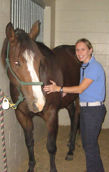
2). Palpation: This is a thorough “hands-on” exam of the horse which may help reveal any heat, swelling, sensitivity, or unevenness between limbs. Your veterinarian will palpate the horse’s neck, back, and all four limbs. During this time, she is feeling the bones and joint spaces, in addition to several soft tissue structures such as muscles, tendons, and ligaments. Your veterinarian may palpate the limbs with the horse standing and with each limb elevated. She will often use hoof testers to evaluate any sole or heel sensitivity.
3). Baseline Lameness: After taking a thorough history and palpating your horse, your veterinarian will establish your horse’s baseline lameness. This is the lameness that is noted at a walk or a trot in a straight line. The grades of lameness, as established by the American Association of Equine Practitioners (AAEP), are defined below:
- Grade 5=non-weight bearing lameness
- Grade 4=consistently lame at the walk
- Grade 3=consistently lame at the trot
- Grade 2=inconsistently lame at the trot/consistently lame with exacerbation
- Grade 1=inconsistently lame under all circumstances
4). Exacerbation of the Lameness: During this part of the exam, your veterinarian will flex your horse’s limbs for a set amount of time, and then watch him move off at a trot in a straight line. Flexion stresses the joints and soft tissues in the region being flexed. The most common flexions are lower front limb, upper front limb, lower hind limb, and upper hind limb. These “flexion tests” are then graded on a scale of 0-3 pending how lame the horse is when he moves off following the test.
Your veterinarian will also watch your horse lunge during this part of the exam. She will watch the horse move at the walk, trot, and canter in both directions. It is important to note if the lameness is worse when the affected limb is on the inside or the outside of the circle. In general, soft tissue lameness is worse on the outside of the circle because the limb is swinging wider, stressing the flexible tissue. Bony/joint lameness is often worse on the inside of the circle because those limbs are bearing more weight.
Surface is typically the final variable to address when trying to exacerbate your horse’s lameness. Whether the lameness improves or worsens on hard vs. soft surface can be an important clue in evaluating lameness.
*It is important to remember that lameness does not always “follow the rules.” Veterinarians utilize lots of general principles when evaluating lameness, but each case is diagnosed and treated specifically.
5). Localizing the Lameness: This involves using a short-acting local anesthetic to desensitize certain areas of the lame limb. Veterinarians typically start “blocking” the nerves that are low on the limb to rule out heel or foot pain. If the horse goes sound after the block is performed, then your veterinarian knows that the lameness is coming from that part of the limb. Joints can also be individually blocked if we suspect that a specific joint is causing lameness.
6). Imaging: After deciding what part of the limb is causing the lameness, your veterinarian will attempt to image the actual lesion. The most common types of imaging are radiography (“X-rays”) and ultrasound. Radiographs best image the bony structures, and ultrasound is typically used for soft tissues. If the lesion cannot be imaged with radiographs or ultrasound, your veterinarian may suggest a referral for nuclear scintigraphy (“bone scan”), MRI, CT, or thermography.
7). Treatment: After establishing the likely cause of your horse’s lameness, your veterinarian will develop a treatment plan that is specific for your horse. Possible treatments include anti-inflammatories, systemic joint support medications, shoeing/trimming recommendations, shockwave therapy, and joint injection with steroids, hyaluronic acid, stem cells, IRAP, or PRP. Your veterinarian will also make rest and/or return-to-work recommendations for you and your horse.
So, when will my horse be sound, Doc? This can be a very difficult question to answer. The type of lesion and your horse’s age will play a role. However, the most important factor is how your horse responds to the treatment plan. Veterinarians often have to revisit the plan pending your horse’s progress. Additional treatment may be needed or the work schedule may need to be changed. Some horses are sound within days, and others continue to be treated for years. It is also very important to take into account your horse’s job. Sometimes we may not be able to get the horse back to his original level of competition (ex. barrel racing, high-level dressage, or eventing), but he may be sound enough for light pleasure riding.
TAKE HOME MESSAGES:
- Every case is different!
- There are lots of diagnostic and treatment options.
- Lameness requires time and patience!
About Dr. Erin Chastain
Dr. Erin Chastain received her Bachelor of Science degree from West Virginia University in 2003. From there, she went on to veterinary school at the University of Georgia. Dr. Chastain graduated in the spring of 2007 and then completed an equine internship at the University of Missouri. For the past 12 years, Dr. Chastain has practiced as an equine veterinarian--six years in south central Pennsylvania and six years in the north Atlanta area. An avid hunter/equitation rider throughout her college years, she fell in love with eventing while in vet school. Dr. Chastain works at Thomasville Animal Hospital in Thomasville, Georgia.
Related Articles
What We're Seeing: How Local Conversations Fill Start Boxes
If you've been on Facebook lately, you've probably had this happen. Someone shares a Starter Trial in your Area's Facebook group with a link to the entry page. A friend comments that they're going. Your trainer tags a few students. Before long, you've clicked the link, looked over the details, an...
US Equestrian Announces Additions to USEF Eventing Pathway Program Lists
US Equestrian is pleased to announce additions to the 2026 USEF High Performance Eventing Programs for both the Elite and Pre-Elite lists. Membership is reviewed twice annually for the Eventing Elite and Pre-Elite Program Lists. For more information about the Pre-Elite and Elite Programs, to view...

U.S. World Championship Team Preps at Tryon and Horse Park of New Jersey
Members of the U.S Eventing Team heading to the upcoming FEI Eventing World Championships in Aachen, Germany from Aug. 10-23 selected the Horse Park of New Jersey and the Tryon International Equestrian Center as their final destinations for the finishing touches ahead of travel overseas last week...