Q&A with Dr. Mathieu Spriet on Equine PET Scans
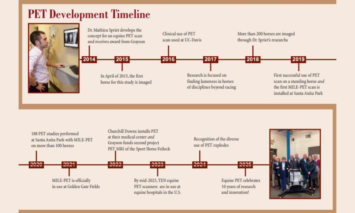
Dr. Mathieu Spriet is an Associate Professor of Diagnostic Imaging at the School of Veterinary Medicine at the University of California- Davis. In 2015, Grayson-Jockey Club Research Foundation funded the first research project that performed positron emission tomography (PET) scans on equine athletes.
What is the process of getting a horse prepared for their PET scan?
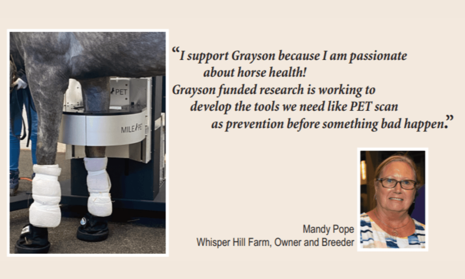
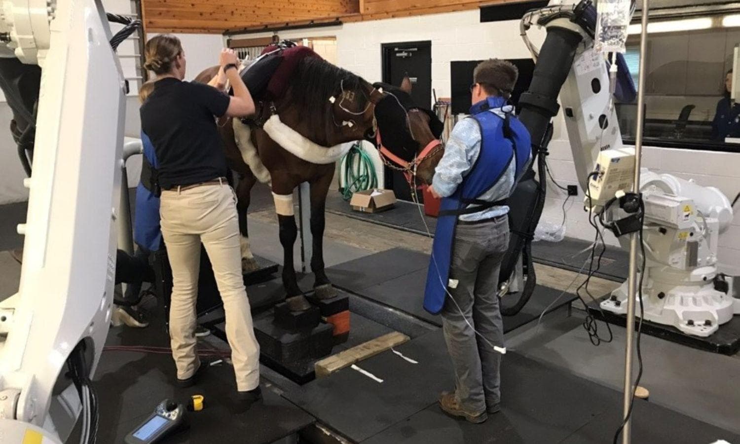
Horses get prepared for a PET scan the same way as for a bone scan. They are injected in a jugular vein via a catheter with a small amount of a radioactive substance, called a radiotracer. For good distribution of the radiotracer to the bones, it is important to have good blood flow to the limbs, making sure the limbs are warm – using wraps and blankets in cold weather is very important. Working the horses on the track the morning of the scan is also helpful. It is necessary to wait 30 minutes to an hour for the radiotracer to leave the bloodstream and get into the bones. Horses can then be sedated, similarly to when performing any other imaging procedure on a standing horse, and images are acquired. Each joint can be imaged in three to five minutes. Horses need to stay in the hospital for a few hours after the scan to clear the radiotracer. Typically, five hours after the initial injection, all radioactivity has disappeared, and the horse can return to a regular barn.
Do you see potential in PET scans being used in other large animal species?
PET is increasingly used in dogs, both for cancer and orthopedic issues assessment. We have imaged a goat for investigation of a lytic bone lesion. In ruminants, there are potential indications for assessment of lameness to rule in or rule out sepsis of the digits, for example. Camelids could potentially be imaged for similar reasons.
Did you see an increase in lameness issues in one breed or discipline over another in your research?
Definitely, based on the breed and discipline we observe different patterns of injuries. If we use the fetlock for example, as this is the joint most commonly imaged with PET, we can see stress remodeling in different areas. Racehorses tend to load more to the back of the joint, and we see common injuries to the palmar aspect of the condyles and sesamoid bones. In sport horses, in particular jumpers, there is more stress on the front of the joint, and the dorsal aspect of the condyles and the proximal phalanx are most commonly the site of injuries.
Interestingly, Quarter Horses in Western disciplines are somewhere in between, showing injuries both at the front and the back of the joint.
Other clinical facts well documented by PET scans:
- Racehorses put a lot of stress on their carpi (knees), with the third carpal bone being most commonly involved.
- Warmbloods and Quarter Horses have more common foot issues, including navicular bone lesions, but also injuries at the attachment of ligaments in the foot.
- Suspensory injuries are recognized across all activities on all breeds.
Do you have any recent success stories where the use of a PET scan helped detect an issue before it became a serious injury?
I cannot mention names, but yes, I can think of a few high-profile horses where PET was instrumental in the decision of retiring to breeding rather than going for an additional season of racing.
But PET does not just stop horses from racing, several horses racing successfully in Triple Crown races and Breeders’ Cup races had been imaged with PET in the weeks to months before the races. One horse was monitored with PET scans over a four-season career, with 15 races and 10 PET scans with strong performances in Breeders’ Cup and international races prior to a healthy retirement.
What is coming down the pike for the future of using PET in the clinic? Will we be able to scan other limbs, e.g., neck and back, eventually?
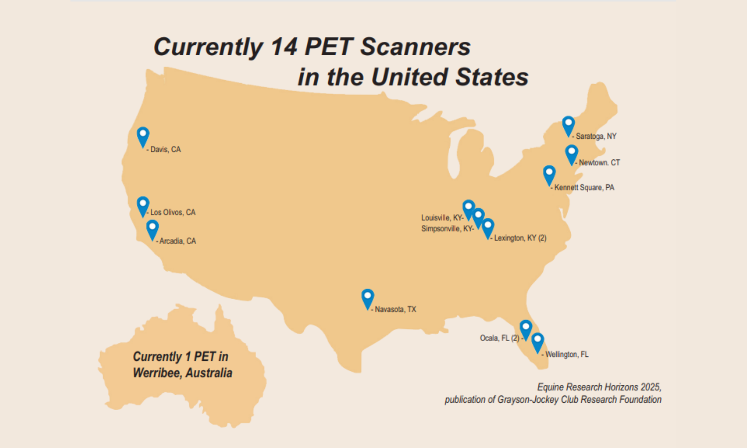
PET scan for horses is becoming increasingly available with 12 active scanners in the U.S., one in Australia, and soon one in Germany. The initial focus was bone imaging in racehorses, but there are now many other applications with Warmbloods and Quarter Horses also commonly imaged. There is also growing interest in soft tissue imaging with the use of a different radiotracer for tendon and ligament imaging.
The scanners and associated software keep improving. The latest generation scanner, that recently was installed at Santa Anita, has additional detectors allowing for scan times twice shorter. Fetlocks can now be imaged in two minutes. Head imaging will occur in the near future, and most likely neck imaging will also become available in a more distant future. These applications obviously require larger scanners to be designed.
Are there any specific types of injuries or conditions for which PET scans provide a major advantage?
PET excels at bone imaging, with the ability to monitor the bone turnover rate. This is particularly pertinent for “subchondral bone” injuries, which are the most common injuries in racehorses and are also increasingly recognized in sports horses. PET is able to recognize when an injury is severe enough that resting is indicated to prevent breakdown, but PET also allows monitoring over time to adjust the training and racing programs accordingly.
Imaging soft tissue with PET is increasingly used in sport horses where it is particularly pertinent regarding suspensory ligament imaging. The suspensory ligament can be difficult to monitor once it has been injured, and the PET soft tissue tracer is helpful in distinguishing mature scar tissue from active injuries.
There are two additional areas where PET is likely to become particularly relevant, these are laminitis and imaging of infection.
Laminitis is a devastating condition, often career ending if not life ending. The underlying causes are extremely complex and involve various triggers of inflammation. PET can provide precise mapping and quantification of the inflammation in the foot, which is extremely helpful for understanding the development of the disease and assessing possible therapeutic options.
Infection in soft tissues, joints, or bones develops secondary to wounds or as a complication of injections or surgical procedures. Early and accurate identification of the infection is key for appropriate treatment. More validation is needed, but PET appears to be able to recognize infection before other imaging techniques.
Grayson-Jockey Club Research Foundation
Grayson-Jockey Club Research Foundation is traditionally the nation's leading source of private funding for equine medical research that benefits all breeds of horses. Since 1940, Grayson has provided nearly $42.3 million to underwrite more than 437 projects at 48 universities. Additional information about the foundation is available at grayson-jockeyclub.org.
Related Articles
Cooling the Threat: How Grayson-Funded Research Is Changing the Fight Against Laminitis
Few diagnoses strike fear into a horse owner's heart like laminitis. This painful and often devastating disease damages the sensitive tissues that connect the hoof wall to the underlying coffin bone. Laminitis not only threatens a horse's athletic career but remains one of the leading causes of p...
Understanding Air Quality Decisions at US Equestrian-Licensed Competitions
As wildfire smoke and other environmental conditions continue to impact air quality in parts of the country, competitors may find themselves wondering how decisions are made regarding whether an equestrian competition proceeds as scheduled, is modified, or is canceled.

Alliston Claims Fourth CCI4*-L Championship and The Event at Rebecca Farm Concludes Its 25th Year
What began as a big idea of Rebecca Broussard’s marked 25 years of having an even bigger impact on hundreds of riders, horses, and the American eventing community.